Project Goal: To define the role of PI3-kinase and Akt in the CSF-1 growthand survival program in myeloid cells
The primary aim of this project is to develop an understanding of how the Colony Stimulating Factor-1 (CSF-1) supports the proliferation and survival of myelomonocytic cells. To do this, we genetically complemented a CSF-1 receptor (CSF-1R) mutant defective in mitogenic and survival signaling with downstream signaling molecules. DKI is a CSF-1R mutant that lacks the receptor binding site for PI3-kinase but can still activate this pathway via an alternate route involving Src family kinases and Gab2. Unlike cells expressing the WT receptor, CSF-1 cannot support the proliferation and survival of DKI cells. CSF-1 induction of the PI3-kinase/Akt pathway in DKI cells was significantly impaired but stable expression of a constitutively active p110* restored their ability to function mitogenically in response to CSF-1. These results establish that direct recruitment of PI3-kinase to the receptor is essential for optimal activation and normal proliferative and survival functions require a threshold level of PI3-kinase activity.
Both Akt-dependent and independent pathways downstream of PI3-kinase played a role as stable expression of a constitutively active Akt (E40K) was unable to completely substitute for p110* in restoring the mitogenic response of DKI cells. DKIcells in the presence of CSF-1 undergo a protracted form of cell death displaying several features characteristic of apoptosis (Fig. 1). Cell death was not accompanied by early changes in delta Psi-m or by an early release of cytochrome c into the cytosol. Atypical also of apoptosis was the relative lack of detectable caspase activation associated with dying DKI cells (Fig. 2). Overexpression of the pro-survival molecule Bcl-xL delayed but did not prevent death of DKI cells in the presence or absence of CSF-1. These results are consistent with a form of cell death that is independent of mitochondrial dysfunction and caspase activation.
See our latest publication on the role of PI3K and Akt in cell survival pdf.
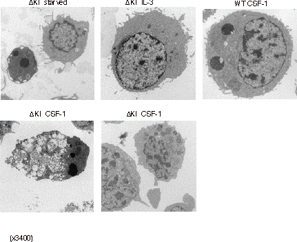
Fig. 1 Electronmicrographs. Starved: deprived of growth factors –typical marginated and condensed chromatin of apoptotic cells. IL-3: cells grown in IL-3 are healthy and nuclei show the fine lacy pattern. WT CSF-1:cells expressing the WT CSF-1R are healthy when grown in CSF-1. DKI CSF-1:cells expressing DKI CSF-1R are dying in the presence of CSF-1: a mix of late apoptotic cells with heavy vacuolization (left) and cells with some early changes (right).
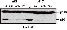
Fig. 2 PARP cleavage shown by immunoblotting. PARP is asubstrate of several executioner caspases and is cleaved into a large fragment (p85) and a small fragment (not shown). Starved cells are included as a positive control for PARP cleavage. Aliquots of cells are processed at the indicated times after CSF-1 treatment.